400 876 6002

Optical coherence tomography (OCT)


| OCT | |
|---|---|
| Methodology | Spectral domain OCT |
| Axial resolution | ≦6um(in tissue) |
| Transverse resolution | ≦20(in tissue) |
| Scan depth | ≥2.5mm(in air) |
| Scan range | 6-12mm |
| Scan speed | ≥24,000A-scan/sec,up to 36,000A-scan/sec |
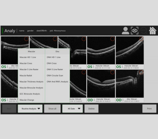
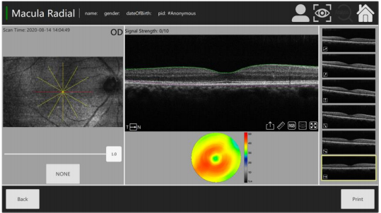
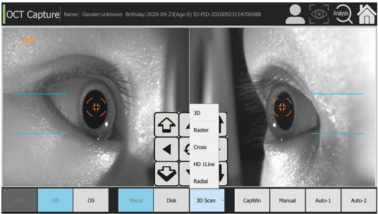
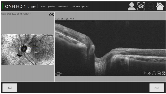
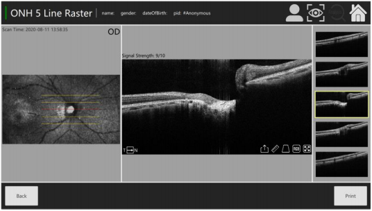
| Scan mode | 3D(Macular&Optic Disk) ,HD,Raster,Circle,Cross |
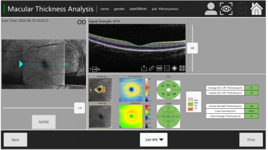
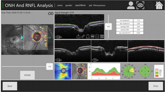
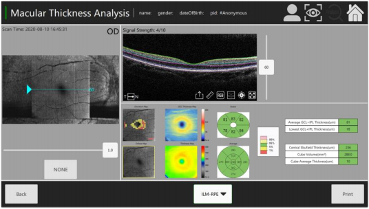
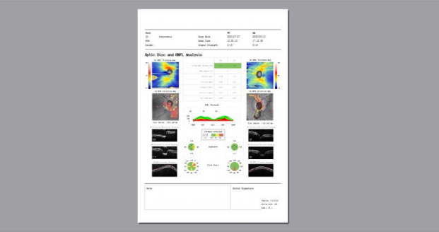
| Analysis mode | Up to 7 retinal layers segmentation,Macular analysis mode,RNFL&Optic Disk analysis mode,Glaucoma analysis mode and Progress analysis for follow-up examination. |
| Fundus image | OCT en face |
| Focus adjustment | -15D to+15D |
| Pupil diameter | ≥3mm |
| OCT light source | 840nm SLD |
| Operation | 13.3inch touchscreen,optional external mouse or keyboard |
| Power supply | 100-240V,50/60HZ |
| Dimensions | 497x395x490mm |
| Weight | 34kg |